A group from Institute of Parasitology, McGill University, Ste-Anne-de-Bellevue, QC, Canada, etc. has reported on glycan profile of Schistoeoma mansoni EVs and those origins.
https://www.mdpi.com/2076-0817/10/11/1401/htm
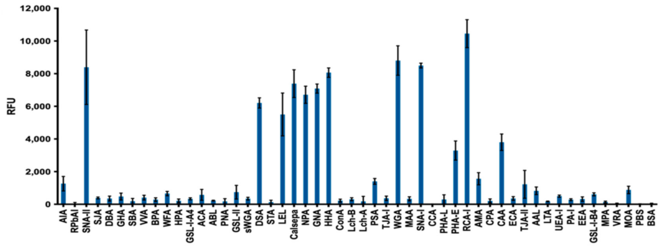
Using lectin microarrays, it was identified several lectins that exhibit strong adhesion to Schistosoma mansoni EVs, suggesting the presence of multiple glycan structures on these vesicles. Interestingly, SNA-I, a lectin that recognises structures with terminal α2-6 sialic acid, displayed strong affinity for S. mansoni EVs. SNA-II and RCA-I, which have affinity for structures which are frequently terminated with sialic acids showed strong intensities as well. In addition, the four mannose-binding lectins, Calsepa, NPA, GNA and HHA, showed strong intensities, suggesting abundance of high-mannose glycans.
Question is the origin of sialic acid.
To identify worm structure(s) as possible sources of EVs, we performed histochemistry on whole adult worms with three lectins that exhibited strong affinity for S. mansoni EVs (DSA, RCA-I and SNA-I). 100% of tegumental cells are positive for SNA-I labelling, DSA labels definitive tegumental cells and the gut of the parasite, and RCA-I labelled various structures within the worm, including potential tegumental cells and what appear to be the excretory pore and excretory tubules. Together, these results may indicate the presence of multiple EV sub-populations and the involvement of the tegument and the digestive and protonephridial (excretory) systems in the release of EVs from adult schistosomes.
This finding on glycosylation profile of Schistosoma mansoni EVs and those origins is of interest, as sialic acids play important roles in the context of infection by aiding immune evasion, affecting target recognition, cell entry, etc.