blog

Changes in glycan modification accompanied by change from ESC to epiblast-like cells (EpiLCs) and its control factor behind them

A group from Soka University etc. has reported on changes in glycan modification accompanied by change from ESC to epiblast-like cells (EpiLCs) and a control factor behind them.
https://www.nature.com/articles/s41598-020-79666-4

With the change from ESC to EpiLCs, the following changes in glycan modification occur:

  1. For N-type glycans, the high mannose structure is the main structure in common, but in EpiLCs, fucose modification, bisecting, and Sia modification are increased, for Sia, α2-6 is the main linkage, and typeI LacNAc (Galβ1-3GlcNAc) is highly expressed with α1-2Fuc modified.
  2. For O-type sugar chains, Tn antigen and O-GlcNAc are the main in common, but in EpiLCs, the expression of O-glycan is generally increasing, and elongated mucin type O-glycans also increase.
  3. For Glycosaminoglycans (GAG), EpiLCs generally increase GAG expression, especially Heparan sulfate (HS), chondroitin sulfate (CS) and dermatan sulfate (DS).
  4. For glycolipids, EpiLCs cause structural changes from globo (Gb) to ganglio (Gg).

Behind these glycan modification, polycomb repressive complex 2 (PRC2), a chromatin protein, was shown to be involved as a regulator. A number of glycosyltransferase genes involved in glycan modification of ESC is under the control of PRC2.
Characteristic changes in glycan modification in high-grade gliomas

A group of Quilmes National University, Bernal, Buenos Aires Province, Argentina has reported characteristic glycan modification in gliomas.
https://www.oncotarget.com/article/27850/text/

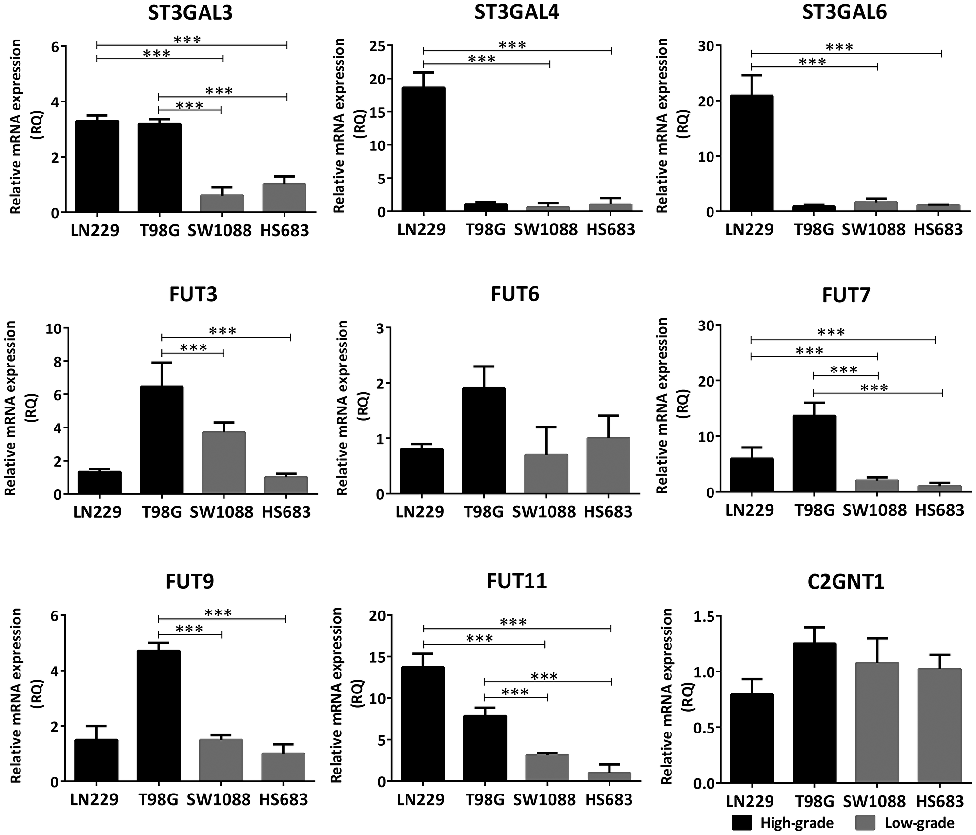
In high-grade gliomas, its glycan modification is characterized by the progression of terminal sialic acid of N-glycans and multi-antennary N-glycans. The sialic acid modification is mainly α2-3 linkage along with the terminal fucosylation, suggesting that the terminal modification structure of N-glycans is SLex. To support this, glycosyltransferases (α1,3-fucosyltransperases (FuT3-7 and 9-11) and α2,3-sialyltransferases (ST3Gal3/4/6) ) are also highly expressed.

They concluded that changes in the glycan profile of tumor cell surfaces are the basis for the development of therapeutic drugs targeting such aberrant glycans and novel glycan markers.


A method using ammonia to select hepatocytes from differentiated ones from pluripotent stem cells (iPSC, ESC)

Toxicology tests using hepatocytes have come to be widely used as an alternative way for testing drug toxicology using animals. The problem with using hepatocytes in toxicology tests is that it is difficult to supply large quantities of hepatocytes with the same characteristics. Therefore, a group from the National Institute of Child Health and Development proposes a method to create hepatocytes from pluripotent stem cells (iPSC, ESC) and enrich highly uniform hepatocellular populations from heterogeneous cell populations using ammonia.
https://www.biorxiv.org/content/10.1101/2020.03.20.999680v1.full

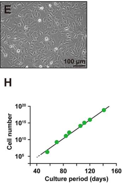
After ESC is differentiated into hepatocytes, an ammonia treatment is performed for 2 days to select uniform hepatocytes that are resistant to ammonia toxicity. 70-80% will die at this stage. Surviving hepatocytes are cultured and proliferated on MEF-feeder. Hepatocytes selected in this way multiply about 30 times in 190 days. The detailed mechanism of ammonia toxicity is not known, but it is thought that ammonia ions compete with potassium ions and eventually die with intracellular and/or extracellular pH change. ALB, AFP, CYP3A4, CPS1, and OTC genes are highly expressed in ammonia-selected hepatocytes, and ALB and AFP decrease with the passage. CPS1 and OTC are genes involved in the metabolism of ammonia. CYP3A4 is one of the enzymes that metabolizes unwanted biological molecules.
The same is possible with hepatocytes made from iPSC.

The differentiated cells from iPSC and /or ECS were further cultivated and propagated in the ESTEM-HE medium (GlycoTechnica, Ltd.) .

Establishment and characteristics of a new immortalized hepatocyte strain, HepaMN

Hepatocytes are essential for toxicology testing of pharmaceuticals in vitro. Hepatocyte strains such as HepG2, Huh7, THLE-2, PLC-PRF-5, and AML-12 are used. A group from the National Center for Child Health and Development has established a new immortalized hepatocytes  named HepaMN from from a liver associated with biliary atresia. Immortalization was performed by inoculation with CDK4, cyclin D1, and TERT.
https://www.nature.com/articles/s41598-020-73992-3

Characteristics of HepaMN strains:
Established from untransformed hepatocytes, and has normal liver function and diploid chromosomes,
Expression of albumin gene at the same level as HepaRG,
Showing the cell morphology of hepatocytes,
Induces stable cytochrome P450 3A4 (CYP3A4) and shows normal metabolic effects,
Efficiently proliferates and is stable even in very long passages,
As a result, succeeded in providing a new and useful hepatocyte strain for toxicology testing.


HepaMN can be effectively cultured by EMUKK-15

Development of vaccines using adeno-associated virus (AAV) against the new coronavirus (SARS-CoV-2)

Existing Pfizer and Modelna vaccines for the new coronavirus (SARS-CoV-2) are mRNA type, but the following group is evaluating prototype vaccines using adeno-associated viral (AAV).
https://www.biorxiv.org/content/10.1101/2021.01.05.422952v3.full.pdf

Therapeutic gene transfer using AAV is recognized as a highly safe method, and Glybera, Luxturna, Zortensma etc. have already been approved by the FDA and EMA as gene therapeutics.
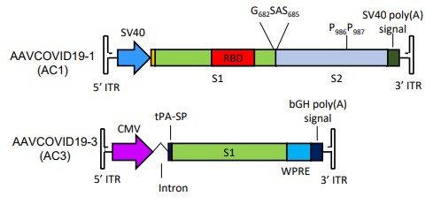
Vaccines using two types of AAV have been prototyped. AAVCOVID-19-1 (AC1) encodes a full-length S protein, which expresses an antigen S protein on the cell membrane of an infected cell. Another, AAVCOVID-19-3 (AC3), encodes part of the S protein, and the antigen takes the form of a secretory protein.


The evaluation was carried out with mice and nonhuman primates, and the immune response was retained for about 5 months with a single doze. There is no problem with storage at room temperature for one month, and vaccines are amenable to large scale production because the methodology has been established. As an effect, AC1 seems to be higher than AC3.

However, the blog Admin has some concerns that the antigen genes are incorporated into cells.

Effects of the new coronavirus (SARS-CoV-2) mutations in the UK and South Africa on a vaccine

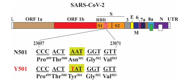
It has been pointed out that mutations in the new coronavirus (SARS-CoV-2) are behind infection explosions in the UK and South Africa. A number of mutations have occurred, but a mutation common to these has already been shown to be N501Y.
A group of The University of Texas Medical Branch, Galveston TX has reported on how the mutation affects the effects of Pfizer’s vaccine, Tozinameran (BNT162b2, a nucleoside modified RNA vaccine).
https://www.ncbi.nlm.nih.gov/pmc/articles/PMC7805448/

A comparative experiment using N501 and Y501, which is the genetic background of N501,  was done by measuring  50% plaque reduction neutralization titers for N501 and Y501 viruses using sera from 20 people collected 2 weeks and 4 weeks after administration of the vaccine. As a result, it was reported that almost the same neutralization titers were obtained.

In the new coronavirus (SARS-CoV-2), heparan sulfate is involved in the capture of viruses, and sialic acid modification of ACE2 weakens viral binding

A group from The University of Hong Kong etc. has reported on the effects of heparan sulfate and glycan modification of ACE2 in the infection of the new coronavirus (SARS-CoV-2).
https://www.nature.com/articles/s41467-020-20457-w

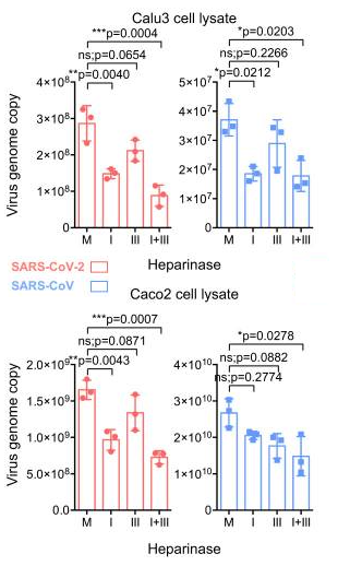
Calu3 (lung epithelial cells) and Caco2 (intestine epithelial cells) are used in SARS-CoV-2 infection experiments. Heparinase was used to investigate the effects of heparan sulfate, and Neuraminidase (NA) was used for the effects of ACE2 glycan modification, with a particular focus on sialic acid.

As shown in the figure below, heparinase suppresses viral infection and shows that heparan sulfate is involved in the capture of the virus as a co-receptor for ACE2. For the sialic acid modification of ACE2, it is shown that the infection of the virus is rather stronger by cleaving the sialic acid with NA.

Evaluation of glycan structure of reference mAb (humanized IgG Type 1) in NIST, U.S.A.

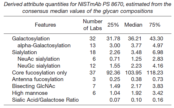
NIST in the U.S. offers humanized IgG Type 1 mAb as a reference mAb. NS0 cells are used to manufacture this mAb. NS0 cell is a model cell line derived from non-secretory mouse myeloma, which is commercially used in biomedical research and the production of therapeutic proteins.
https://pubmed.ncbi.nlm.nih.gov/31591262/

The status of glycan addition is reported by comprehensively following the results of 103 evaluations conducted at 73 institutions around the world. It is likely to be useful as glycan modification information of reference mAb of IgG Type 1.

Priority of vaccine administration for the new coronavirus (COVID-19): Should over-60s still be prioritized?

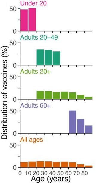
A group from The University of Colorado Boulder etc.  has simulated on the priority of vaccine administration for the new coronavirus (COVID-19).
https://www.ncbi.nlm.nih.gov/pmc/articles/PMC7743091/

Five age groups of vaccination were assumed, and simulated changing the combination of the following parameters: vaccine rollout speeds to the population (0.05% to 1%/day), infection rates (1.15, 1.5), vaccine efficacy (90%), and also the case that the effectiveness of the vaccine decreases with age (60 years old = 90% – > 80 years old = 50%).
The results vary depending on the conditions, and the best choice replaced between the case that prioritizes the over-60s and the one that prioritizes 20 ~ 59 year-olds. However, since the prerequisites are always changing in reality, overall, it would be a better choice to prioritize the over-60s .

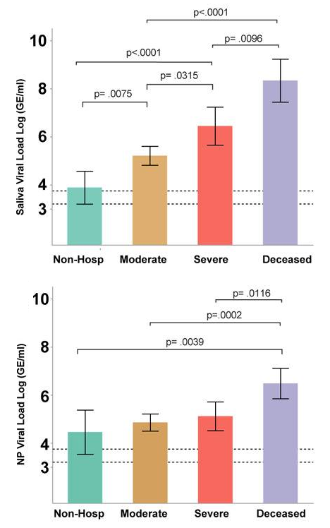
The most dynamic unifying predictor of the disease pathology of the new coronavirus (COVID-19) is the saliva viral load.

A group from Yale University School of Medicine etc. has reported that the most dynamic unifying indicator of the pathophysiology of the new coronavirus (COVID-19) is the saliva viral load.
https://www.ncbi.nlm.nih.gov/pmc/articles/PMC7805468/

Existing markers for COVID-19 include inflammatory cytokines and chemokines (CXCL10, IL-6, IL-10), inflammasome (IL-18, IL-1β), interferon (IFNα, IFNγ, IFNλ), etc.), however, they have found that the saliva virus load was very well correlated with the disease pathology. The nasopharyngeal viral load was also evaluated as a comparison. The amount of virus in saliva and nasopharyngeal (RNA copies/mL) was calculated from Ct values of RT-PCR extracting RNA from saliva. For the discrimination accuracy among non-hospitalization, moderate, severe, and deceased, strong predictive ability was obtained as follows, moderate disease (AUC = 0.96), severe disease (AUC = 0.89), and fatal (AUC = 0.91).

The upper figure of the below shows the correlation between the saliva viral load and the disease pathology, and the lower figure shows the nasopharyngeal viral load and the disease pathology.

Powered by WordPress |Copyright © 2020 Emukk. All rights reserved