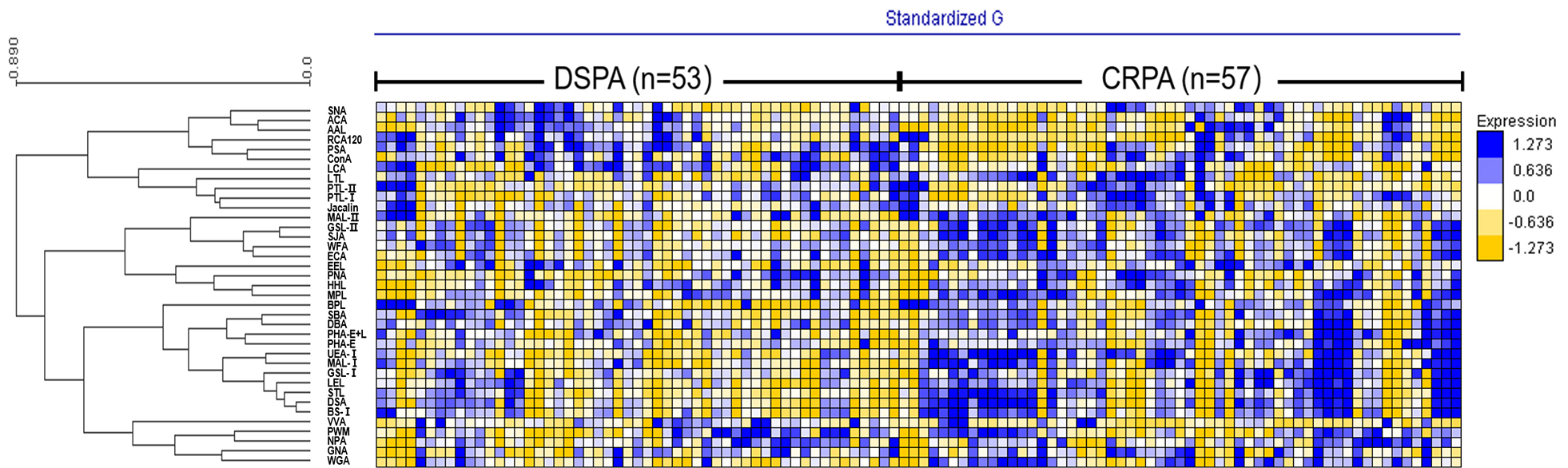
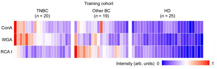
Detailed N-glycan analysis combining Glycan profiles taken by Lectin Microarrays and AI

A group from Department of Bioengineering, University of California, San Diego, La Jolla, CA 92093, USA, etc. has reported a lectin and AI-based approach to predict N-glycan structures and determine their relative abundance in purified proteins based on lectin-binding patterns.
https://www.biorxiv.org/content/10.1101/2024.03.27.587044v1

This method can be used when the number of glycanss to be evaluated is limited, but there are a lot of problems when applying it generally.

A similar software named “SA/DL easy” had been created by Mx using Deep Learning as a core technology 5 years ago. By using this software, you can quickly do the same thing.
The problem lies in the tedious work of creating training data, or preparing a large number of expressed glycan structures whose structures have been properly identified, and obtaining glycan profiles.
https://www.emukk.com/SADL-Easy_Eng/index.html

Detecting Triple-Negative Breast Cancer with using Glycan Profiling of Extracellular Vesicles

A group from Beijing Engineering Research Center for BioNanotechnology, CAS Key Laboratory of Standardization and Measurement for Nanotechnology, National Center for Nanoscience and Technology, Beijing, China, etc. has reported about glycan profiling of extracellular vesicles (EVs) for detecting triple-negative breast cancer (TNBC).
https://www.ncbi.nlm.nih.gov/pmc/articles/PMC10937950/

A panel of 3 lectins (ConA, WGA and RCA I) was used to detect the EV surface glycan profiles unique to TNBC.
As a result, they succeeded in getting an area under ROC curve (AUC) of 0.91 with using the weighted sum of 3 lectins (ConA, WGA and RCA I) for discriminatiing TNBC from other BCs and HDs.

<a?

They say that prostate cancer can be detected by using its exosomes, but

A group from Institute of Chemistry, Slovak Academy of Sciences, Bratislava, etc. has reported about a measurement using a sandwich scheme with CD63, exosomes and SNA lectin for detecting prostate cancer.
https://www.ncbi.nlm.nih.gov/pmc/articles/PMC10892626/

In this experment, exosomes produced by prostate cancer cells are examined as a new biomarker for detecting prostate cancer.

Comparing with exosomes produced by benign (control) cell line RWPE1 and carcinoma cell line 22Rv1, it was showen that
(1) the control exosomes mainly interacted with SNA and MAAII lectins; however, they exhibited a lower affinity than the carcinoma exosomes, and also
(2) PHA-L and PHA-E were only able to bind poorly to control-derived exosomes, while there were no interactions to carcinoma exosomes.
This result is quite reasonable because usually the signal intensity of PHA-L and PHA-E disapper with fully sialylated N-glycans suggesting that sialylation is stronger in carcinoma exsosomes than that of control exosomes.

However, blog author is skeptical about their conclusion that it is possible to perform measurements in a sandwich configuration, i.e., antibody/exosomes/lectin, because exsosmes are generally strongly sialylated and CD63 can not discriminate exsosomes produced by prostate cancer cells from other exosomes.

VVA Lectin characteristically binds to invasive urothelial carcinomas

A group from Department of Urology, Gifu University Graduate School of Medicine, Gifu, Japan, etc. has reported about characteristic glycan marker in invasive urothelial carcinomas.
https://www.ncbi.nlm.nih.gov/pmc/articles/PMC10806140/

The study found that a specific lectin, VVL, was present in cases of invasive urothelial carcinoma and its variant components. More intense VVL staining was observed with invasive or muscle invasive urothelial carcinomas and urothelial carcinomas with variant components than that in non-invasive urothelial carcinomas

VVL recognizes the GalNAc residue linked to serine or threonine in a polypeptide Tn antigen. Other glycan structures, such as Galβ1,3GalNAc-α-Ser/Thr (T antigen) and GlcNAcα1,6-GalNAc-α-Ser/Thr, including terminal α1,4- and β1,4-linked GalNAc, were also recognized by VVL, but with a weaker affinity.

VVA will have the potential to serve as a promising target for drug delivery in future clinical studies.

The paper has been published: FDA announced High-throughput Glycan Profiling Analysis with a 9-Lectin Microarray for Therapeutic IgG1 mAbs

This paper regarding FDA’s dedicated lectin microarray (14-well lectin microarray using 9 kinds of lectins) for the evaluation of mAb drugs (IgG1) has been published.
This paper is related to the Mx blog post on Dec. 8th, 2023.
https://www.tandfonline.com/doi/epdf/10.1080/19420862.2024.2304268?needAccess=true

The 9 kinds of lectins used in this paper and those glycan binding specificities are summarized as follows.
rPhoSL -> core fucose
PHAE -> bisecting GlcNAc
PHAL -> tri/tetra antennary
MAL_I -> α2-3Sia
rPSL1a -> α2-6Sia
RCA120 -> β-Gal
rOTH3 -> terminal GlcNAc
rMan2 -> high mannose
rMOA -> α-Gal
Note that lectins with an “r” at the beginning of their name indicate that they are recombinant.
rOTH3, rMan2 are not official names.
Contact the author of this blog to learn what the official name is.

Pregnancy diagnosis targeting to changes in urinary glycopatterns

A group from Shaanxi Key Laboratory for Animal Conservation, College of Life Sciences, Northwest University, China, etc. has reported about a possibility of pregnancy diagnosis targeting changes in urinary glycopatterns.
https://www.ncbi.nlm.nih.gov/pmc/articles/PMC10783609/

This study investigates the urinary glycopatterns of golden snub-nosed monkeys (GSM) with using lectin microarrays. It was shown that the types, amounts and structure of N-glycans and the proportion of sialylation and fucoslation of N-glycans are different between pregnant and non-pregnant females, and between (non-pregnant) females and males. This method will provide reference information for pregnancy diagnosis and sex identification, which will benefit the management of the animals.

where, pregnant (P) and non-pregnant (NP) females, and females (F) and males (M)

FDA announced High-throughput Glycan Profiling Analysis with a 9-Lectin Microarray for Therapeutic IgG1 mAbs

To evaluate glycan epitopes of therapeutic IgG1 mAbs, FDA has developed a new lectin microarray with 9 kinds of lectins, and has demonstrated its effectiveness for high-throughput glycan profiling analysis using GlycoStation Reader 2300 (GSR2300) .
https://www.fda.gov/media/169026/download
2023 FDA Science Forum

The new lectin microarray (IgG1-mAb-LecChip) developed by FDA immobilizes 9 kinds of lectins: rPhosL, rOTH3, RCA120, rMan2, MAL_I, rPSL1a, PHAE, rMOA, and PHEL, and uses a standard 14 wells LecChip format.
Glycan analysis of IgG1 mAbs can be performed using lectin microarrays without creaving glycans, making it possible to perform high-throughput glycan profiling analysis from intact IgG1.
FDA has recommended pharmaceutical companies to use IgG1-mAb-LecChip and GlycoStation to facilitate high-throughput glycan profiling analysis when developing IgG1 mAbs to assess batch-to-batch or biosimilar-to-innovator glycan epitopes.

The figure below shows how IgG1-mAb-LecChip, which was optimized for IgG1 glycan analysis, was developed using GlycoStation and LecChip (n=74 library).

As an example of showing the effectiveness of this technology, the figure below shows the result of evaluating the differences in glycosylation between Infliximab and its biosimilar using IgG1-mAB-LecChip and GSR2300. It can be clearly seen that there are significant differences in the abundance of High Mannose structure, sialic acid modification, and triantennary N-glycans.

PAA-glycans are used to target cancer cells, and cytotoxic oxygen radicals are released by light irradiation to selectively kill cancer cells

A group from School of Chemistry, University of East Anglia, Norwich Research Park, Norwich, UK, etc. has reported about a new DDS using glycan-lectin interactions and photodynamic therapy.
https://pubs.rsc.org/en/content/articlelanding/2023/NA/D3NA00544E

Lectin expression can be altered in diseased-state cells.
For example, in cancer the glycan–protein interactions play key roles in avoiding immunosurveillance and reattachment to new tissue during metastasis.
Cancer cells are also associated with increased metabolism due to their unregulated, increased growth, reflected in an increase of glycan transporters.
For breast cancer cells, there are key lectins and glycan-binding receptors that are upregulated, which include galectins, glucose transporters, and the mannose receptor.

PAA-glycans and an amine derivate of the photosensitiser chlorin e6 were chemically attached onto Au nanoparticles as a new DDS.
PAA-glycans act as targeting molecules onto the target cancer cells, and the photosensitiser releases cytotoxic reactive oxygen species upon activation with light of a specific wavelength to kill cancer cells.

Methods to promote colonization and activation of Bacillu. in the rhizosphere: SynCom and Prebiotics

A group from College of Resources and Environmental Sciences, Nanjing Agricultural University, Nanjing, China etc. has reviewed about plant biocontrol mechanisms of Bacillus.
https://ami-journals.onlinelibrary.wiley.com/doi/10.1111/1751-7915.14348

As is well known, species of the genus Bacillus have been widely used for the biocontrol of plant diseases in the demand for sustainable agricultural development.

The “cry for help” mechanism in plant means that plants fight pathogen attack by assembling health-promoting beneficial microbes by releaseing specific signals. This mechanism is very similar to human’s immunity that immune cells secrete cytokines/chemokines, and thereby recruit immune cells further and activate immunity.
The root exudates are extremely crucial for recruiting biocontrol agents (i.e., beneficial microbes like Bacillus) in response to plant diseases, and it has been known that L-malic acid, citric acid, fumaric acid, and tryptophan, threonic acid, lysine, pectin, xylan, and arabinogalactan are key exudates.

The use of Bacillus strains for the biocontrol of plant disease has achieved certain benefits worldwide. However, practical utilization of Bacillus is usually confronted with unstable disease suppression efficacy under field conditions. That is because complicated and dynamic factors, such as soil characteristics, plant genotypes, and indigenous microbiota, can all influence the colonization and functional efficacy of inoculated Bacillus agents.

To overcome this issue, two types of methods have been proposed.
One is to use a method called “SynCom” which is to use a bacterial consortium constructed by using some keystone strains from the genus of Bacillus, Burkholderia, Enterobacter, Lysobacter, Stenotrophomonas, Pseudoxanthomonas, Pseudomonas, and Acinetobacter.
The other is to use “Prebiotics”. As mentioed above, specific signals released from root exudates recruit Bacillus strains and induce their activities. Therefore, relevant compounds can be developed as prebiotics for enhancing root colonization and biocontrol performance, similar to those widely applied for stimulating beneficial bacteria in the human gut. So, exogenous addition of sucrose, L-glutamic acid, riboflavin could be used as prebiotics to promote rhizosphere colonization by beneficial Bacullus strains.

Comparative glycan profiling between drug-sensitive Pseudomonas aeruginosa strains and drug-resistant strains

A group from Laboratory of Functional Glycomics, College of Life Sciences, Northwest University, Xi’an, Shaanxi, China, etc. has reported about comparative glycan profiling between drug-sensitive Pseudomonas aeruginosa (DSPA) strains and carbapenem-resistant Pseudomonas aeruginosa (CRPA) strains.
https://pubmed.ncbi.nlm.nih.gov/37861315/

lectin microarrays were used to analyze the differences in glycan alterations between 53 drug-sensitive DSPA strains and 57 carbapenem-resistant CRPA strains obtained from clinical isolates, with the goal of identifying important glycopatterns associated with carbapenem resistance.

In this experiment, whole bacterial cell lysates, which were fluorescently labeled with Cy3, were applied onto Lectin microarrays to take glycan profiles of bacteria.

As a result, it was found that LCA could be an strong biomarker detecting differential expression of glycan structures on the bacterial surface between DSPA with CRPA.