ACE2 and BSG/CD147 are involved in SARS-CoV-2 infection of human iPS cell-derived podocytes

A group from Department of Biomedical Engineering, Pratt School of Engineering, Duke University, Durham, NC, USA, etc. has reported that ACE2 and BSG/CD147 are involved in SARS-CoV-2 infection of human iPS cell-derived podocytes.
https://pubmed.ncbi.nlm.nih.gov/34816259/

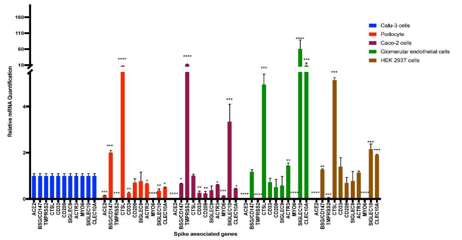
Human iPS cell-derived podocytes express many host factor genes (including ACE2, BSG/CD147, TMPRSS2, CTSL, CD33, DC-SIGN/CD209, SIGLEC9, SIGLEC10, ACTR3, CLEC10A) associated with SARS-CoV-2 binding and viral processing. It is known that proteases such as Transmembrane Serine Protease 2 (TMPRSS2) or cathepsin L (CTSL) promote fusion and internalization of the receptorviral spike complex. Human iPS cell-derived podocytes expressed lower levels of ACE2 and TMPRSS2 when compared to Calu-3, and both BSG/CD147 and CTSL were expressed at high levels as shown below.

Nevertheless, podocytes derived from human iPS cells were directly infected with SARS-CoV-2 at low Multiplicity of Infection (MOIs) of 0.01 to 1, and intriguingly, there was significantly more viral uptake in the podocytes than Calu3 (p-value < 0.0001) and Caco-2 cells (p-value < 0.0001). Pre-treatment of infected podocytes with both anti-ACE2 and anti-BSG/CD147 antibodies at a concentration as low as 0.1 µg/ml significantly diminished viral uptake (p-value < 0.0001), suggesting that both ACE2 and BSG/CD147 are involved in SARS-CoV2 internalization in human iPS cell-derived podocytes.

Plants detoxify Tetrabromobisphenol A (TBBPA) by glycosylation, but rhizobacteria deglycosylate it again.

A group from Research Center for Eco-Environmental Sciences, Chinese Academy of Sciences, Beijing, China, etc. has reported that plants detoxify Tetrabromobisphenol A (TBBPA) by glycosylation, but rhizobacteria deglycosylate it again.
https://www.ncbi.nlm.nih.gov/pmc/articles/PMC8603600/

TBBPA is among the most commonly used brominated flame retardants owing to its excellent properties and low price, and its annual application is continually increasing.
Plants can absorb TBBPA from water, soil, and air. Plants can transform various organic contaminants. The biotransformation of organic compounds in plants can be categorized into phase I, II, and III reactions according to the green liver concept of phytoremediation. For phenolic compounds that contain active hydroxyl groups, glycosylation is an important and common phase II reaction.

Glycosylated metabolites are generally more water soluble than parent contaminants and are therefore easily excreted from plant roots to the outside solutions. Some studies have reported that glycosylated metabolites can also be bound to cell walls and/or compartmentalized in special cell organelles, such as vacuoles. Thus, the formation of glycosylated metabolites can prevent contaminants from accumulation and translocation within plants, and they can decrease the toxic effects of the contaminants on plants.

In this work, the formation of glycosylated metabolites of TBBPA in pumpkin plants and their subsequent excretion from roots to hydroponic solutions effectively decreased the accumulation of TBBPA in plants. However, the deglycosylation of glycosylated metabolites mediated by the microorganisms back to their parent TBBPA revealed more complex uptake and biotransformation processes in these plant–microorganism systems. The TBBPA produced from the observed deglycosylation process can be then absorbed and cause adverse effects on plants and other soil organisms again.

Survival times of SARS-CoV-2 and Influenza virus on three types of postcards with different surface coating

A group from Graduate School of Medical Science, Kyoto Prefectural University of Medicine, Kyoto, Japan, etc. has reported stability of SARS-CoV-2 and Influenza virus on three types of postcards with different surface coating.
https://www.ncbi.nlm.nih.gov/pmc/articles/PMC8590490/

The assessment of the risk of virus transmission through papers, such as postcards, is important. However, the stability of severe acute respiratory syndrome coronavirus 2 (SARS-CoV-2) and influenza A virus (IAV) on different types of papers is currently unknown.

In this experiment, stability of SARS-CoV-2 and IAV was evaluated on the surface of postcards with three types of coatings, plain paper (PP), inkjet paper (IP), and inkjet photo paper (IPP).
The survival times of SARS-CoV-2 and IAV were significantly shorter on IP and IPP than on PP as shown below (SARS-CoV-2). Therefore, switching from PP to IP or IPP is effective in reducing the viral transmission risks through paper.

Fungal cell wall polysaccharides β-(1, 3)-glucan and α-(1, 3)-glucan activate the Wnt/β-catenin pathway in human DCs.

A group from Institut National de la Santé et de la Recherche Médicale, Centre de Recherché des Cordeliers, Sorbonne Université, Université de Paris, France, etc. has reported that C-type lectins are necessary in induction of the Wnt/β-catenin pathway and fungal cell wall polysaccharides β-(1, 3)-glucan and α-(1, 3)-glucan, but not chitin, activate the Wnt/β-catenin pathway in human DCs.
https://journals.asm.org/doi/10.1128/mBio.02824-21

Aspergillus fumigatus is an omnipresent airborne fungal pathogen. Although inhaled conidia are usually eliminated in healthy individuals, they may cause hypersensitization, severe asthma with fungal sensitization, allergic bronchopulmonary aspergillosis, colonization of altered respiratory epithelium, and aspergilloma in existing pulmonary lesions.

Innate immune cells, including macrophages, dendritic cells (DCs), and neutrophils, are involved in antifungal activity against A. fumigatus. Upon fungal encounter, DCs engage their various pattern recognition receptors (PRRs) to recognize the evading pathogen. These A. fumigatus-educated DCs subsequently instruct distinct CD4+ T-cell polarization like Th1, Th2, Th17, and FoxP3+ regulatory T cells (Tregs). Among these, Th2 and Th17 responses are nonprotective for Aspergillus infection. On the other hand, Th1 cells have a major role for the induction of protective immune responses. Although Tregs are immunosuppressive and promote chronic and persistent infection, they are also critical for preventing inflammation-associated tissue damage. Therefore, the balance between Th1 and Treg responses is critical for the protective immune response against A. fumigatus.

Recent studies have demonstrated the involvement of Wnt/β-catenin pathway for the induction of tolerogenic functions in DCs and promotion of Treg responses via various anti-inflammatory mechanisms, like expression of IL-10, transforming growth factor beta (TGF-β), and retinoic acid.

By using A. fumigatus as a model, it was shown that fungal species activate the Wnt/β-catenin pathway in human DCs, along with the secretion of Wnt ligands Wnt1 and Wnt7a. Inhibition of the Wnt pathway resulted in decreased DC maturation and selective inhibition of anti-inflammatory cytokine IL-10 without affecting the secretion of most of the proinflammatory cytokines. Abrogation of the Wnt/β-catenin pathway in DCs also led to reduced Treg polarization without altering the polarization of other CD4+ T-cell subsets.

It was also found that C-type lectins are involved in induction of the β-catenin pathway and A. fumigatus’s cell wall polysaccharides β-(1, 3)-glucan and α-(1, 3)-glucan, but not chitin, activate the β-catenin pathway in human DCs.

where, CA=unstimulated, SC=stimulated with swollen conidia

Cortical-bone-derived stem cells (mCBSCs) treatments effectively improve myocardial structure and functions after myocardial infarction compared to MSC: Difference in glycosylation

A group from Research Team for Geriatric Medicine (Vascular Medicine), Tokyo Metropolitan Institute of Gerontology, Japan, etc. has reported that cortical-bone-derived stem cells (mCBSCs) treatments effectively improve myocardial structure and functions after myocardial infarction compared to MSC.
https://www.ncbi.nlm.nih.gov/pmc/articles/PMC8584423/

Recently, it have been shown that mouse cortical-bone-derived stem cells (mCBSCs) improve cardiac remodeling and functions. The mCBSC-treated hearts showed increased neovascularization, and newly formed cardiac myocytes were also observed. mCBSCs produce a unique combination of immunomodulatory and angiogenic and proangiogenic factors, which may be the reason why mCBSCs were more effective in improving the post-myocardial infarction hearts compared to cardiac-derived stem cells and MSCs.

The self-renewing ability in mCBSCs was higher than that in mMSCs, but the mCBSCs exhibited only chondrogenic differentiation, while the mMSCs exhibited adipogenic, osteogenic, and chondrogenic differentiation. It was also found that mCBSCs secrete a greater amount of TGF-β1 compared to mMSCs, and that the TGF-β1 contributed to the self-migration of mCBSCs and activation of fibroblasts. Migrated CBSCs expressing TGF-β1 may contribute to converting cardiac fibroblasts into myofibroblasts at an infarcted site.

As for cell surface glycans, the relative intensities of three lectins WFA (lacdiNAc), ECA (lactosamine-binding lectin), and MAL-I (α2-3 sialic acid binding lectin) in mCBSCs were higher than those in mMSCs. Moreover, the relative intensities of the three lectins, SNA, SSA, and TJA-I (α2-6 sialic acid binding lectins), in mCBSCs were significantly lower than those in mMSCs. It was considered that this lower expression of α2-6sialic acid may be an indication of specific differentiation towards chondrogenic lineage and not towards adipogenic or osteogenic differentiation.

Previous research has shown that glycans contribute to regulation of the signaling mediated by leukemia inhibitory factor (LIF), Wnt, FGF, bone morphogenetic protein BMP, and Notch, which are required for the maintenance of stem cells. It has been also known that WFA-binding glycans on LIF receptorβ and gp130 regulate LIF/STAT3 signaling, which is required for self-renewal of mouse embryonic stem cells.

In this study, it was found that WFA-binding glycans are more specific to CBSCs than MSCs, and this suggested that WFA-binding glycans may contribute to the self-renewal of CBSCs by regulating LIF/STAT3 signaling, although further studies is required.

Lactobacillus agilis isolated from rhizosphere shows potential as a novel biotherapeutic agent

A group from Department of Biosciences, COMSATS University Islamabad (CUI), Islamabad, Pakistan, etc. has reported that Lactobacillus agilis isolated from rhizosphere of the medicinal plants Ocimum tenuiflorum, Azadirachta indica, Ficus carica shows potential as a novel biotherapeutic agent.
https://www.ncbi.nlm.nih.gov/pmc/articles/PMC8568817/

Potential probiotic bacteria can be used as a biotherapeutic agent and a sustainable alternative to antibiotics, as an anti-oxidative, anti-inflammatory, and anti-diabetic agent without causing any serious side effects. In this experiment, the following assays were performed to select potentials.

Safe status assays
First of all, to exclude potentially harmful bacterial strains, testing of safe status was done before selection of bacterial strains for human as probiotics.
(1) should be negative for blood hemolytic activities and (2) did not show a breakdown of gelatin.

Antibiotic susceptibility
The strains which show less or no resistance to antibiotics were selected, and L. agilis was the best.

In-vitro antibacterial assay
The antibacterial results showed the significant marked antagonistic activity of L. agilis NMCC-15 to kill or inhibit the growth of all the tested bacterial pathogens (Escherichia coli, Pseudomonas aeruginosa, Staphylococcus aureus, Listeria monocytogenes and Bacillus cereus).

Antioxidant, anti-diabetic, and anti-inflammatory assays
Percentage inhibition of the free radical from the mean values showed the comparable effects of L. agilis supernatant to the standard drug with values 68% and 73%, respectively.
L. agilis inhibited the activities of the α-amylase (anti-diabetic potential) by 51.3% than control.
The supernatant of L. agilis showed 61.6% for denaturation albumin protein while aspirin showed 69% activity.

The most abundant bacteria and fungi in seedlings rhizobacteria came from their seeds, not the soil

A group from MaxPlanck Tandem Group in Plant Microbial Ecology, Universidad del Valle, Cali, Colombia, etc. has reported that the most abundant bacteria and fungi in seedlings rhizobacteria came from their seeds, not the soil.
https://www.frontiersin.org/articles/10.3389/fmicb.2021.737616/full

Until recently, scientists have traditionally believed that all the rhizosphere microbiome “is recruited from the main reservoir of microorganisms present in soil. However, Is it really true?

In this experiment, plants were grown in hermetically sealed jars on sterile sand as a way to observe microbiome development in the absence of any other source of inoculum except seeds. In this experiment, 18 kinds of plant were evaluated.

Amazingly enough, in this experiment it was confirmed that soil contributes to microbial diversity in the rhizosphere, however the most abundant bacteria and fungi in rhizospheres derive from seeds. Soil significantly increased bacterial diversity in rhizospheres, however, the highest read abundance was of seed-transmitted bacteria. Only an average of 26% of bacterial taxonomic units (OTUs) came from seeds, but these were responsible for an average of 72% of the reads. These OTUs were mostly Proteobacteria of the genera Pantoea, Enterobacter, Pseudomonas, and Massilia, which were observed in seeds or spermospheres and have also been observed associated with a variety of plant seeds.

Compared to bacteria, there was less seed-transmitted fungal diversity in rhizospheres, with only an average of 12%; however, these OTUs tended to become abundant, representing an average of 42% of the reads. Of these seed-transmitted rhizospheric fungi, Fusarium was the most abundant, occurring in all soil-grown rhizospheres, as it did in all spermospheres.


In this experiment, unsterilized seeds were planted in either sterile sand or farm soil inside hermetically sealed jars. The sand was sterilized by autoclaving twice for 20 min at 121°C, and after transfer to glass jars, it was autoclaved a third time for 20 min at 121°C, and the soil was excavated from a fallow cassava field at a CIAT property near Palmira, Colombia.

Changes in N-glycosylation on neuroblastoma affect on cell proliferation and invasion

A group from Department of Biochemistry and Molecular Biology, East Carolina University, Greenville, North Carolina, USA, etc. has reported that changes in N-glycosylation on neuroblastoma affect on cell proliferation and invasion.
https://journals.plos.org/plosone/article?id=10.1371/journal.pone.0259743

Two types of neuroblastoma cells were used. One is an ​engineered N-glycosylation mutant cell line with Mgat1 knockdown, NB_1(-Mgat1), expressed solely oligomannose N-glycans, and the other was its parental cell line, NB_1 expressed significant levels of higher order N-glycans, complex and hybrid N-glycans as shown below (GNL and ConA have higher affinity to oligomannose type N-glycans, and E-PHA and L-PHA have higher affinity to complex type N-glycans).

Cell growth was faster in NB_1 cells with complex type N-glycans than in NB_1(-Mgat1) cells without complex type N-glycans. Quantitatively speaking, there was about a 54% decrease in cell proliferation between NB_1(-Mgat1) cells and NB_1 cells.

NB_1(-Mgat1) cells had more and longer protrusions than NB_1 cells, indicating that the glycosylation mutant cells solely expressing oligomannose type N-glycans were more invasive than those expressing complex types of N-glycans. To quantify cell invasiveness, the ratio of measured invasive area to the measured sphere area was measured. The cell invasiveness of the NB_1(-Mgat1) cell line was 2.3-fold greater than the NB_1 cell line.

Two types of multivalent lectins produced from hemagglutinins of clostridium botulinum: Gg binds to Gal/GalNAc and Rn binds to Neu5Ac

A group from Tokyo University of Agriculture and Technology, Tokyo, 183-8509, Japan, etc. has reported two types of multivalent lectins produced from hemagglutinins of clostridium botulinum.
https://www.nature.com/articles/s41598-021-01501-1

Clostridium botulinum is a gram-positive anaerobic bacterium that produces a neurotoxin. Botulinum bacteria are classified into serotypes A to G depending on the antigenicity of the toxin produced. The type C 16S progenitor toxin is composed of a neurotoxin, non-toxic non-hemagglutinin component and several hemagglutinin proteins (HA) designated as HA1, HA2, HA3a and HA3b.

It has been know that type C HA1 has two binding sites: site I and site II. Site I binds to N-acetylneuraminic acid (Neu5Ac), N-acetylgalactosamine (GalNAc) and galactose (Gal) while site II binds to galactose only.

Authors have developed two new multivalent lectins from the above mentioned hemagglutinins of clostridium botulinum designed to have different binding specificities were produced: namely Gg and Rn:
Gg is an Alexa 488 labeled complex with HA1 WADF -HA2 WT-HA3 WT, which binds to Gal/GalNAc,

and, Rn is an Alexa 594 labeled complex with HA1 NQAA-HA2 WT-HA3 WT, which binds to Neu5Ac only.

Auto-antibodies against AngII were developed with SARS-CoV-2 infection

A group from Pritzker School for Molecular Engineering, University of Chicago, Chicago, Illinois, USA, etc. has reported that auto-antibodies against AngII were developed with SARS-CoV-2 infection.
https://www.ncbi.nlm.nih.gov/pmc/articles/PMC8575143/

Surprisingly, it was found that a substantial proportion, 63% (73/115), of the COVID-19 patients had positive levels of anti-AngII autoantibodies, where 115 were hospitalized COVID-19 patients convalescent from a SARS-CoV-2 infection.

First, it was pointed out that the levels of anti-AngII were significantly higher in patients with dysregulated blood pressure as shown below.

Further, the effect of anti-AngII autoantibodies on oximetric saturation (SF ratio) was measured. The lowest daily mean SF ratio was compared between the COVID-19 patients who developed auto-antibodies against AngII (73 patients) and those who did not (42 patients) as shown below.

It was considered that SARS-CoV-2 and AngII to ACE-2 might lead to their co-phagocytosis by antigenpresenting cells, thus providing a strong immune adjuvant (the virus molecules) to the self-peptide AngII, leading to an anti-AngII autoimmune response.